If the course is stable, visit frequency is gradually reduced.
Key points#
This quick reference answers three practical questions: how high the risk of graft disease is, how often the patient should be examined, and what to do at the first signs of an adverse course.
Principle 1Risk determines follow-up intensity
Vascularization, repeat PK, active infectious inflammation, severe dry eye, and systemic disease place the patient in the high-risk group.
Principle 2Assess the same core findings at every visit
Visual acuity, graft clarity and epithelialization, suture status, keratic precipitates/edema, inflammation, new vessels, and IOP.
Principle 3Taper topical corticosteroids slowly
Abrupt unsupervised discontinuation may trigger graft rejection. In high-risk eyes, maintenance treatment may be prolonged or lifelong.
Principle 4Deterioration requires an immediate decision
Redness, photophobia, pain, blurred or reduced vision require urgent slit-lamp examination and escalation—not waiting for scheduled follow-up.
Critical error
Do not “observe over time” suspected rejection, infection, wound/suture failure, or graft melt without urgent corneal surgeon review.
What to assess at every visit#
Use a reproducible minimum examination set to detect subtle changes before irreversible graft opacity develops.
Function
Uncorrected and corrected visual acuity; ask about blur, halos, photophobia, and pain.
Graft
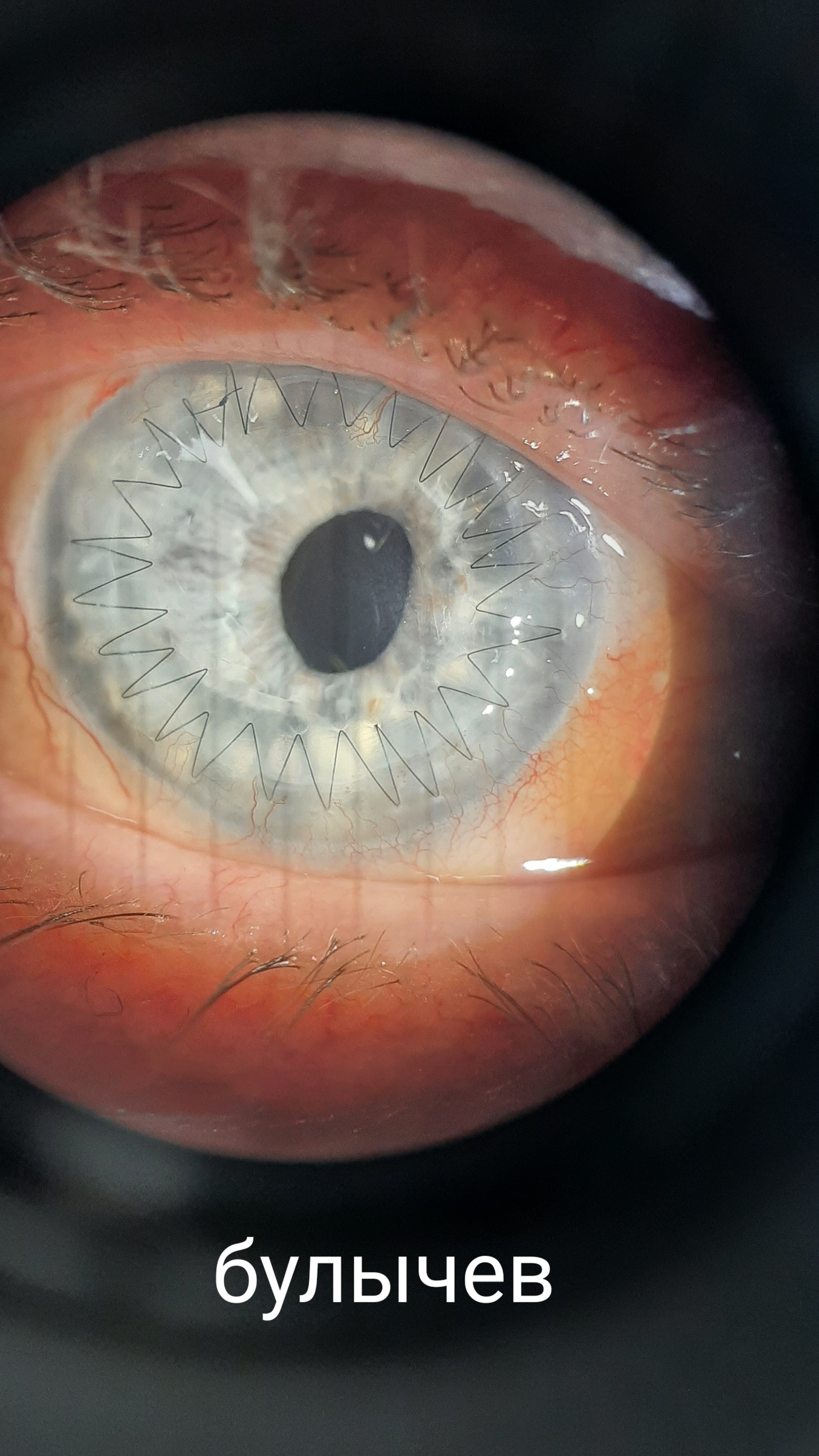
Clarity, completeness of epithelialization, stromal edema, Descemet membrane folds, keratic precipitates, and focal infiltrates.
Suture and wound
Tension or loosening, exposed suture, erosion, infiltrate, vessel ingrowth; perform a Seidel test if leakage is suspected.
Inflammation and infection
Injection pattern, anterior chamber cells/flare, hypopyon, discharge, and epithelial defects.
Intraocular pressure
Check at every visit. After PK, non-contact tonometry may be unreliable; the source document proposes transpalpebral tonometry or comparative digital palpation.
Additional tests
Keratometry/topography at approximately month 3; after suture removal, repeat refraction and topography in 2–4 weeks.
Uncomplicated course: epithelialization is usually complete by days 7–10; edema gradually decreases over 1–2 months. By month 6, the expected findings are a quiet eye, clear graft, clean intact suture, and no significant vascular invasion.
Immunologic risk stratification#
Low risk
Favorable recipient bed and first elective optical graft.
- First PK.
- Avascular cornea or only minimal superficial vascularization.
- No active inflammation.
- Typical indications: keratoconus, corneal dystrophies, limited stromal opacities.
- No severe systemic autoimmune disease.
High risk
Corneal immune privilege is disrupted or healing is substantially impaired.
- Corneal vascularization; involvement of ≥2 quadrants indicates very high risk.
- Repeat keratoplasty.
- Urgent PK for ulceration, perforation, or melt.
- Severe dry eye, Sjögren syndrome, or ocular cicatricial pemphigoid.
- Autoimmune disease, systemic vasculitis, or Stevens–Johnson syndrome.
- Combined surgery, pediatric age, or poor-quality donor tissue.
High risk requires a low threshold for referral
A new vessel reaching the graft, increasing opacity, edema, thinning, or marked inflammation → urgent corneal surgeon review.
Follow-up schedule#
Low risk#
- 1 week after discharge
Approximately 2–3 weeks after surgery: visual acuity, slit-lamp examination, tonometry, suture assessment, and adherence review.
- First month
Examine every 2 weeks.
- Months 2–6
Approximately monthly; by month 3, perform keratometry or topography if epithelialization is complete.
- Months 6–12
Every 1.5–2 months, or at least once per quarter.
- After year 1
During year 2, every 3–6 months; thereafter annually for life.
Sutures: with a favorable course, sutures are generally left untouched for at least 12 months. Removal is usually considered at 12–15 months, individualized by clarity, vascularization, and wound strength.
High risk#
- 3–4 days after discharge
First outpatient review.
- First month
Every 3–7 days; if unstable, every other day during the first 2 weeks.
- Months 2–6
Every 2 weeks, at least twice monthly.
- Months 6–12
Monthly.
- Year 2 and beyond
At least every 2 months during year 2; thereafter quarterly for life.
Escalation and referral
In high-risk eyes, rejection, marked inflammation, a new vessel, thinning, or melt should not be managed for a prolonged period in outpatient care alone—refer to a specialist center.
Baseline treatment: low risk#
The doses and intervals below are retained from the methodological recommendations. Adjust treatment according to the operating surgeon’s discharge instructions and clinical course.
Scroll horizontally to view the table
| Medication / regimen | Months 1–2 | Months 3–4 | Months 5–6 | Months 7–8 | Months 9–10 | Months 11–12 |
|---|---|---|---|---|---|---|
| Antibiotic | 1 drop × 4 times/day** ONLY 7–10 DAYS | — | — | — | — | — |
| Corticosteroid (dexamethasone 0.1%) | 1 drop × 6 times/day | 1 drop × 5 times/day | 1 drop × 4 times/day | 1 drop × 3 times/day | 1 drop × 2 times/day | 1 drop once daily (morning) |
| Corneal surface protective agent | 1 drop × 3 times/day | — | — | 1 drop × 2 times/day | — | — |
| Lubricant | 1 drop × 4–8 times/day | As needed; preservative-free preferred | ||||
* drops × times per day; ** course ONLY 7–10 DAYS.
The source document provides two early dexamethasone regimens
Table regimen: dexamethasone 0.1%, 1 drop 6 times daily during months 1–2. Narrative regimen: every 2 hours while awake or 6–8 times/day. Do not independently average these regimens; follow the surgeon’s discharge instructions and graft status.
Antibacterial prophylaxis
- Agents
- Ciprofloxacin 0.3%, ofloxacin 0.3%, levofloxacin 0.5%, or tobramycin 0.3%.
- Regimen
- 4–6 times daily. Course: ONLY 7–10 DAYS.
- Discontinue
- After epithelialization if there are no signs of infection.
Lubrication
- Regimen
- 4–8 times daily as needed.
- Preferred
- Preservative-free products.
- Goal
- Support epithelialization and treat postoperative dry eye.
Mydriatics / cycloplegics
- Agents
- Tropicamide 1% or cyclopentolate 1%.
- Regimen
- 1–2 times daily during the first 1–2 weeks — USE WITH EXTREME CAUTION AND ONLY ON THE OPERATING SURGEON’S RECOMMENDATION.
- Monitoring
- IOP monitoring is mandatory; consider the risk of Urrets–Zavalia syndrome. Use only on the operating surgeon’s recommendation.
EXTREME CAUTION: only on the operating surgeon’s recommendation.
IOP-lowering treatment
- First line
- Timolol 0.5% twice daily or dorzolamide 2% 2–3 times daily.
- Caution
- Prostaglandin analogues are generally avoided during the first months.
- Approach
- Do not automatically stop corticosteroids: balance the risks of ocular hypertension and graft rejection.
Treatment when indicated#
- Herpetic etiology: valacyclovir 1000 mg orally 3 times daily for 10 days, or famciclovir 500 mg 3 times daily. Topical option: acyclovir 3% ointment at night.
- Topical NSAIDs: diclofenac 0.1% or bromfenac 0.05% once daily during the first weeks. Not mandatory; use cautiously because of delayed epithelialization and epithelial erosion.
- Systemic corticosteroids: not routinely required. In some centers, prednisolone approximately 20–30 mg with tapering over 2–3 weeks.
Treatment: high risk#
Intensive topical corticosteroids
Dexamethasone 0.1% hourly for at least 2 weeks → 6 times/day by month 2 → 4 times/day by month 3 → 2–4 times/day through month 6 and beyond.
Long-term maintenance
Do not discontinue completely during the first 1–2 years; lifelong once-daily maintenance may be required.
Cyclosporine A 0.05%
In selected cases: 1 drop twice daily for at least 6–12 months, often up to 2 years; only when prescribed by the surgeon and etiologically appropriate.
Ocular surface support
Lubricants every 1–2 hours, gels/ointments at night; consider punctal plugs or partial tarsorrhaphy when indicated.
Progressive vascularization
Consult a corneal transplant center; local anti-VEGF therapy is an individualized option based on multidisciplinary review.
Infectious-disease caution
If fungal infection is possible or bacterial susceptibility data are unavailable, do not introduce topical corticosteroids or cyclosporine A until the pathogen is clarified and adequate antimicrobial control is established.
Refractory rejection: prednisolone 1 mg/kg orally + cyclosporine A 5 mg/kg orally if not contraindicated; alternatively tacrolimus or mycophenolate mofetil—only under specialist supervision.
Acute deterioration: differential management#
First identify the dominant syndrome: immune rejection, infection, mechanical failure, epithelial defect, or graft melt. Then begin permissible initial measures and urgently escalate care.
Scroll horizontally to view the table
| Situation | Key findings | Initial action | Escalation and referral |
|---|---|---|---|
| Rejection | Reduced vision, redness, photophobia; edema, Descemet membrane folds, keratic precipitates, Khodadoust line. | Dexamethasone 0.1% hourly; every ~2 hours overnight. | Urgent referral to a specialist center / hospital. |
| Suture-related keratitis | Focal infiltrate and erosion around the suture, discharge, progressive ulceration. | Intensive topical fluoroquinolone therapy; temporarily stop corticosteroids until infection is controlled. | Urgent review; remove/replace the suture if the infiltrate fails to resolve within 2–3 days. |
| Suture / wound failure | Positive Seidel test, shallow anterior chamber, wound deformation, history of trauma. | Bandage contact lens, sterile eye dressing, supine face-up position, frequent antibacterial drops. | Immediate transfer for resuturing. |
| Persistent epithelial defect | Fluorescein-staining defect, pain, absent epithelialization, filaments. | Frequent lubricants, carbomer at night, therapeutic soft contact lens; temporarily hold corticosteroids for a significant defect. | If persistent for >2 weeks, investigate dry eye, infection, and mechanical causes. |
| Graft melt | Progressive thinning, graft deformation, impending or established perforation. | Stop corticosteroids and initiate intensive corneal surface protective therapy. | Urgently consider autoconjunctival corneal coverage, blepharorrhaphy, or repeat large-diameter keratoplasty. |
Graft rejection#
TopicalDexamethasone 0.1% hourly; every ~2 hours overnight
Hospital treatmentDexamethasone 16 mg IV or prednisolone 60–80 mg/day with gradual taper
Suture failure or wound dehiscence#
Emergency
Bandage contact lens + sterile dressing + supine face-up position + frequent antibacterial drops → immediate transfer for resuturing.
Suture-related keratitis and infection#
- Antibiotic: intensive topical fluoroquinolone therapy.
- Suture: if the infiltrate does not resolve within 2–3 days, remove or replace the infected suture.
- Corticosteroids: temporarily discontinue until infection is controlled.
Persistent epithelial defect#
- Frequent artificial tears; carbomer at night.
- Therapeutic soft contact lens.
- Consider autologous serum tears.
- For a significant defect, temporarily hold corticosteroids until epithelialization.
- If the defect persists for more than 2 weeks → investigate dry eye, infection, and mechanical causes.
Graft thinning and melt#
Threat to globe integrity
Stop corticosteroids, initiate intensive corneal surface protective therapy, and urgently consider autoconjunctival corneal coverage, blepharorrhaphy, or repeat large-diameter keratoplasty.
Late follow-up and additional procedures#
Scroll horizontally to view the table
| Clinical task | Practical approach | Approximate timing |
|---|---|---|
| Suture removal | Individualize according to clarity, vascularization, suture type, and wound strength. Repeat refraction/topography after removal. | Usually 12–15 months; review 2–4 weeks after removal. |
| Post-keratoplasty astigmatism | Spectacle correction; rigid gas-permeable lenses if the epithelium is stable. Excimer laser correction only with a stable graft. | RGP lenses after 3–4 months; laser correction no earlier than 2 years. |
| Cataract | Perform phacoemulsification + IOL under protective corticosteroid therapy. | Preferably 1–2 years after PK. |
| Refractory glaucoma | Laser/surgical treatment when indicated, accounting for the risk of triggering rejection. | If possible, after graft healing—6–12 months. |
| Repeat optical PK | Completely control inflammation and minimize vascularization; decide jointly with the corneal surgeon. | No earlier than 6–12 months after the first graft. |
Critical errors#
- Abruptly stop topical corticosteroids or allow the patient to change dosing frequency independently.
- Use corticosteroids or cyclosporine A when fungal infection is possible or antimicrobial control is inadequate.
- Ignore a new vessel, focal infiltrate near a suture, or subtle reduction in graft clarity.
- Rely solely on non-contact tonometry after PK without correlating it with clinical findings and an alternative IOP assessment.
- Continue prolonged outpatient treatment of suspected rejection, wound failure, or melt without urgent escalation.
- Plan refractive or repeat surgery before the graft is stable and inflammation is controlled.
Prepared by#

References#
The primary source is the authors’ methodological recommendation; the bibliography is retained in a concise clinical format.
- Arzhimatova GSh, Salikhov EA, Chernakova GM, Shemyakin MY. Algorithms for management of patients with a full-thickness corneal graft in Moscow public healthcare organizations during outpatient care. Methodological recommendations. Moscow: Moscow City Ophthalmology Center, S.P. Botkin Center; 2025.
- Eye Bank Association of America. Eye Banking Statistical Report — 2024.
- Eye Bank Association of America. 2018 Eye Banking Statistical Report. Washington, DC: EBAA; 2019.
- Arzhimatova GSh, Salikhov EA, Shemyakin MY. Corneal neovascularization: current view of molecular mechanisms and therapeutic approaches. Russian Ophthalmological Journal. 2023;16(2):153–159. doi:10.21516/2072-0076-2023-16-2-153-159.
- Sarezky D, Orlin SE, Pan W, VanderBeek BL. Trends in Corneal Transplantation in Keratoconus. Cornea. 2017;36(2):131–137. doi:10.1097/ICO.0000000000001083.
- Boisjoly HM, Tourigny R, Bazin R, et al. Risk factors of corneal graft failure. Ophthalmology. 1993;100(11):1728–1735. doi:10.1016/S0161-6420(93)31409-0.
- Sugar A, Tanner JP, Dontchev M, et al. Recipient risk factors for graft failure in the Cornea Donor Study. Ophthalmology. 2009;116(6):1023–1028. doi:10.1016/j.ophtha.2008.12.050.
- Lois N, Kowal VO, Cohen EJ, et al. Indications for penetrating keratoplasty and associated procedures, 1989–1995. Cornea. 1997;16(6):623–629.
- Gurnani B, et al. Corneal Graft Rejection. StatPearls. 2023. NCBI Bookshelf: NBK519043.
- Magovern M, et al. Trends in keratoplasty for keratoconus: a report by the Eye Bank Association of America. Ophthalmology. 2018;125(10):1471–1478.
- Sharif W, et al. Corneal graft rejection: incidence, features, risk factors, and outcomes. Clinical Ophthalmology. 2019;13:1343–1355.
- Gurnani B, et al. Penetrating Keratoplasty. StatPearls. 2023. NCBI Bookshelf: NBK592388.
- Arzhimatova GSh, Chernakova GM, Salikhov EA, Shemyakin MY. Statistical analysis of risk factors for penetrating corneal graft disease in high-risk keratoplasty. Ophthalmology in Russia. 2024;21(3):509–516. doi:10.18008/1816-5095-2024-3-509-516.
- Slonimskiy YuB, Arzhimatova GSh, Slonimskiy AYu, Salikhov EA, Slonimskaya AS. Penetrating corneal graft survival: analysis of clinical cases. The EYE GLAZ. 2025;27(3):222–230. doi:10.33791/2222-4408-2025-3-222-230.
- Kalinnikov YuYu, Izmailova SB, Ragimova LF, et al. Clinical and functional results of combined surgical treatment of keratoconus: 10-year follow-up. Ophthalmology in Russia. 2025;22(1):191–199. doi:10.18008/1816-5095-2025-1-191-199.
Purpose of this materialThis quick reference is intended for rapid professional orientation and selection of a management pathway. It does not replace the operating surgeon’s individualized decision, discharge instructions, or in-person assessment.